Data compression makes the heart grow fuzzy
By Kimberly Patch, Technology Research NewsAlthough manual compression works well for restarting a heart, it looks like compressing digital angiograms makes it more difficult to diagnose heart problems.
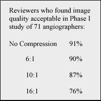
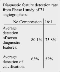
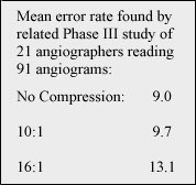
According to a trio of studies done by international researchers, physicians make a few more mistakes reading digital angiograms when they are compressed 10 to 1 than with uncompressed files, and substantially more mistakes when the compression rises to 16 to 1. (See Charts)
Radiologists use compression for static files like x-rays, which tend to average 20 MB uncompressed. Digital coronary angiograms, which are moving x-ray images that show a heart working, are much larger files, averaging 500-600 MB for an 80-second exam. They also require a 7.5 megabits-per-second data rate to accurately display the heart moving in real-time. This makes them prime candidates for compression; shrinking the size of the file would reduce both the data rate needed to see it in real-time and the time needed to transmit it to another location.
Compressing these files, however, is probably a bad idea said Richard Kerensky, who headed one of the three studies, which were all published the April issue of the journal of the American College of Cardiology.
"We found that moderate levels of compression -- 10 to 1 or 16 to 1 -- tended to introduce some errors into an examination," said Kerensky, an associate professor of medicine in the division of cardiovascular medicine at the University of Florida's college of medicine.
"As the compression gets over 10 to 1 ... you might not detect calcium or see a thrombosis in an artery." Kerensky said. This is because the "haziness to an edge" that indicates calcium buildup, or the shadowy look of a thrombosis, or blood clot, are difficult to see even in uncompressed images, said Kerensky. "Even stints we place can be hard to see -- these are small arteries that are in motion."
In the Kerensky study 71 angiogram readers examined 100 angiograms that were compressed at various rates for the following seven diagnostic features: complex stenosis/filling defect, calcification, stints, dissection, bypass grafts, right coronary injections and left coronary injections. The study used lossy JPEG compression on 512 x 512 x 8-bit coronary angiograms and was funded by the American College of Cardiology and the European Society of Cardiology.
Timeline: Not Applicable
Funding: Private
TRN Categories: Applied Computing; Data Structures and Algorithms
Story Type: News
Related Elements: Chart 1, Chart 2, Chart 3
Advertisements:
June 21, 2000
Page One
Nature nurtures nanotech
Virtual physicals at hand
Sandia speeds microtube chip making
Data compression makes the heart grow fuzzy
Multicast promises lighter wireless Internet
News:
Research News Roundup
Research Watch blog
Features:
View from the High Ground Q&A
How It Works
RSS Feeds:
News
Ad links:
Buy an ad link
| Advertisements:
|
 |
Ad links: Clear History
Buy an ad link
|
TRN
Newswire and Headline Feeds for Web sites
|
© Copyright Technology Research News, LLC 2000-2006. All rights reserved.